Cholesterol-regulated cellular stiffness may enhance evasion of NK cell-mediated cytotoxicity in gastric cancer stem cells

Gastric cancer has a high rate of recurrence, and as such, immunotherapy strategies are being investigated as a potential therapeutic strategy. *

Although the involvement of immune checkpoints in immunotherapy is well studied, biomechanical cues, such as target cell stiffness, have not yet been subject to the same level of investigation. *

Changes in the cholesterol content of the cell membrane directly influence tumor cell stiffness. *

In the article “Cholesterol-regulated cellular stiffness may enhance evasion of NK cell-mediated cytotoxicity in gastric cancer stem cells” Lijuan Zhu and Hongjin Wang investigate the effect of cholesterol on NK cell-mediated killing of gastric cancer stem-like cells. *

They report that surviving tumor cells with stem-like properties elevated cholesterol metabolism to evade NK cell cytotoxicity. *

Inhibition of cholesterol metabolism enhances NK cell-mediated killing of gastric cancer stem-like cells, highlighting a potential avenue for improving immunotherapy efficacy. *

This study suggests a possible effect of cancer cell stiffness on immune evasion and offers insights into enhancing immunotherapeutic strategies against tumors. *

Measurement of cell stiffness by AFM:

A customized commercially available atomic force microscope (AFM) and NanoWorld Pyrex-Nitride PNP-TR AFM probes were used for the measurement of cell stiffness by AFM. *

Atomic force microscopy cell stiffness was measured according to standard methodology. *

AFM force curves were captured with a customized AFM placed atop an inverted optical microscope that had a heating stage for live-cell imaging and a ×20 objective. Using an XY stage, the materials were moved until the desired cell, which could be observed under an optical microscope, was positioned beneath the AFM tip. Using a NanoWorld PNP-TR-B AFM cantilever (NanoWorld), the force curves on the cell were collected at a rate of ~ 5 μm·s−1 in the relative trigger mode (15 nm trigger threshold). *

By utilizing a thermal tuning and the deflection sensitivity of 170 nm·V−1, the AFM cantilever spring constant was determined to be 0.08 N·m−1. *

Single cells were measured both before and after treatment at 37 °C. The force curves were processed using the AFM’s analysis software, which also computed the Young’s modulus of the sample. This was accomplished by fitting the approach curve to an indentation of less than 500 nm (to account for stiffness) and assuming a cortical Poisson’s ratio of 0.3.*
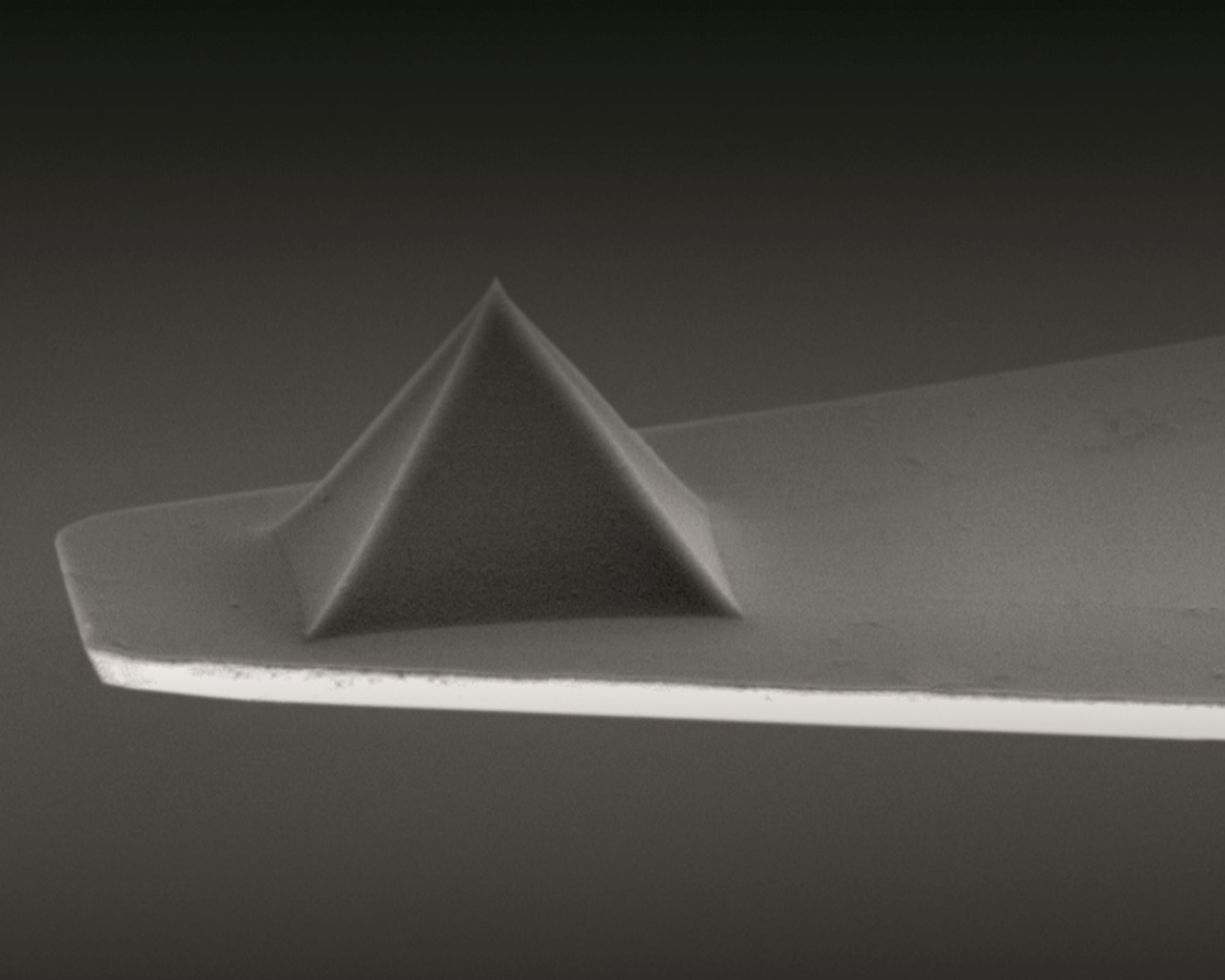
NanoWorld Pyrex-Nitride PNP AFM probe - silicon nitride AFM cantilever and silicon nitride AFM tip
NanoWorld Pyrex-Nitride AFM probe series – AFM tip and AFM cantilever made of silicon nitride

*Lijuan Zhu and Hongjin Wang
Cholesterol-regulated cellular stiffness may enhance evasion of NK cell-mediated cytotoxicity in gastric cancer stem cells
FEBS Open Bio, Volume 14, Issue 5, May 2024, Pages 855-866
DOI: https://doi.org/10.1002/2211-5463.13793

Open Access  The article “Cholesterol-regulated cellular stiffness may enhance evasion of NK cell-mediated cytotoxicity in gastric cancer stem cells” by Lijuan Zhu and Hongjin Wang is licensed under a Creative Commons Attribution 4.0 International License, which permits use, sharing, adaptation, distribution and reproduction in any medium or format, as long as you give appropriate credit to the original author(s) and the source, provide a link to the Creative Commons license, and indicate if changes were made. The images or other third party material in this article are included in the article’s Creative Commons license, unless indicated otherwise in a credit line to the material. If material is not included in the article’s Creative Commons license and your intended use is not permitted by statutory regulation or exceeds the permitted use, you will need to obtain permission directly from the copyright holder. To view a copy of this license, visit http://creativecommons.org/licenses/by/4.0/.

MACC1-Induced Collective Migration Is Promoted by Proliferation Rather Than Single Cell Biomechanics

High metastasis-associated in colon cancer 1 (MACC1) expression is associated with metastasis, tumor cell migration, and increased proliferation in colorectal cancer. Tumors with high MACC1 expression show a worse prognosis and higher invasion into neighboring structures. However, the mediation of the pro-migratory effects is still a matter of investigation.*

In their study “MACC1-Induced Collective Migration Is Promoted by Proliferation Rather Than Single Cell Biomechanics”  Tim Hohmann, Urszula Hohmann, Mathias Dahlmann,  Dennis Kobelt, Ulrike Stein and Faramarz Dehghani aim to elucidate the impact of single cell biomechanics and proliferation on MACC1-dependent migration.*

The authors found that MACC1 expression associated with increased collective migration, caused by increased proliferation, and no changes in single cell biomechanics. Thus, targeting proliferation in high-MACC1-expressing tumors may offer additional effects on cell migration.*

The mechanical properties of single cells were assessed in the form of the Young’s modulus and cortex tension; both were measured using atomic force microscopy. Briefly, cells were seeded on a petri dish and measured 15 min after seeding to avoid slippage of individual cells. Measurements were conducted using a tipless NanoWorld Arrow-TL2 AFM cantilever array to apply a force of 1 nN that led to deformations of 1–2 µm. The Young’s modulus was calculated using the Hertz model.*

NanoWorld tipless Arrow-TL2 AFM probe array with two tipless AFM cantilevers
NanoWorld® Arrow™ TL2 AFM probes are tipless AFM cantilevers for special applications. They can for example be used for attaching spheres and other objects to the free end of the AFM cantilever, or for functionalizing and sensing applications.
The Arrow™ TL2 probes are optionally available with a sample facing side gold coating (Arrow™ TL2Au).
Figure 1 from “MACC1-Induced Collective Migration Is Promoted by Proliferation Rather Than Single Cell Biomechanics” by Tim Hohmann et al. Single cell properties of high- and low-MACC1-expressing colon carcinoma cells. (A,B) depict the results of the biomechanical measurements for the Young´s modulus and the cortex tension. (C,D) show the results of live cell imaging of single cells for the mean speed and the contact area with the substrate. Sample sizes: (A) nSW480/EV = 35; nSW480/MACC1 = 33; nSW620/shMACC1 = 40; nSW620/shCTL = 40. (B) nSW480/EV = 33; nSW480/MACC1 = 31; nSW620/shMACC1 = 25; nSW620/shCTL = 26. (C,D) nSW480/EV = 66; nSW480/MACC1 = 98; nSW620/shMACC1 = 102; nSW620/shCTL = 111. Asterisk depicts statistically significant results with p < 0.05. Box plots show the median (red line), 25 and 75 percentile (box), non-outlier range (whiskers), and outliers (red dots). The mechanical properties of single cells were assessed in the form of the Young’s modulus and cortex tension; both were measured using atomic force microscopy. Briefly, cells were seeded on a petri dish and measured 15 min after seeding to avoid slippage of individual cells. Measurements were conducted using a NanoWorld Arrow-TL2 AFM cantilever to apply a force of 1 nN that led to deformations of 1–2 µm. The Young’s modulus was calculated using the Hertz model.
Figure 1 from “MACC1-Induced Collective Migration Is Promoted by Proliferation Rather Than Single Cell Biomechanics” by Tim Hohmann et al.
Single cell properties of high- and low-MACC1-expressing colon carcinoma cells. (A,B) depict the results of the biomechanical measurements for the Young´s modulus and the cortex tension. (C,D) show the results of live cell imaging of single cells for the mean speed and the contact area with the substrate. Sample sizes: (A) nSW480/EV = 35; nSW480/MACC1 = 33; nSW620/shMACC1 = 40; nSW620/shCTL = 40. (B) nSW480/EV = 33; nSW480/MACC1 = 31; nSW620/shMACC1 = 25; nSW620/shCTL = 26. (C,D) nSW480/EV = 66; nSW480/MACC1 = 98; nSW620/shMACC1 = 102; nSW620/shCTL = 111. Asterisk depicts statistically significant results with p < 0.05. Box plots show the median (red line), 25 and 75 percentile (box), non-outlier range (whiskers), and outliers (red dots).

*Tim Hohmann, Urszula Hohmann, Mathias Dahlmann,  Dennis Kobelt, Ulrike Stein and Faramarz Dehghani
MACC1-Induced Collective Migration Is Promoted by Proliferation Rather Than Single Cell Biomechanics
Cancers 2022, 14(12), 2857
DOI: https://doi.org/10.3390/cancers14122857

Open Access

The article “MACC1-Induced Collective Migration Is Promoted by Proliferation Rather Than Single Cell Biomechanics” by Tim Hohmann, Urszula Hohmann, Mathias Dahlmann,  Dennis Kobelt, Ulrike Stein and Faramarz Dehghani is licensed under a Creative Commons Attribution 4.0 International License, which permits use, sharing, adaptation, distribution and reproduction in any medium or format, as long as you give appropriate credit to the original author(s) and the source, provide a link to the Creative Commons license, and indicate if changes were made. The images or other third party material in this article are included in the article’s Creative Commons license, unless indicated otherwise in a credit line to the material. If material is not included in the article’s Creative Commons license and your intended use is not permitted by statutory regulation or exceeds the permitted use, you will need to obtain permission directly from the copyright holder. To view a copy of this license, visit https://creativecommons.org/licenses/by/4.0/.