Lateral Piezoelectricity of Alzheimer‘s Aβ Aggregates

Alzheimer’s disease (AD) is the most frequent neurodegenerative disorder in the elderly aged over 65.*

The extracellular accumulation of beta-amyloid (Aβ) aggregates in the brain is considered as the major event worsening the AD symptoms, but its underlying reason has remained unclear.

In the article „Lateral Piezoelectricity of Alzheimer‘s Aβ Aggregates“ by Jinhyeong Jang, Soyun Joo, Jiwon Yeom, Yonghan Jo, Jingshu Zhang, Seungbum Hong and Chan Beum Park the piezoelectric characteristics of Aβ aggregates are revealed.

The vector piezoresponse force microscopy (PFM) analysis results exhibit that Aβ fibrils have spiraling piezoelectric domains along the length and a lateral piezoelectric constant of 44.1 pC N-1. Also, the continuous sideband Kelvin probe force microscopy (KPFM) images display that the increment of charge-induced surface potential on a single Aβ fibril is allowed to reach above +1700 mV in response to applied forces.

These findings shed light on the peculiar mechano-electrical surface properties of pathological Aβ fibrils that exceed those of normal body components.*

Both KPFM and nanoindentation measurements were performed using a chemically inert conductive diamond AFM tip. (NanoWorld Pointprobe CDT-FMR).

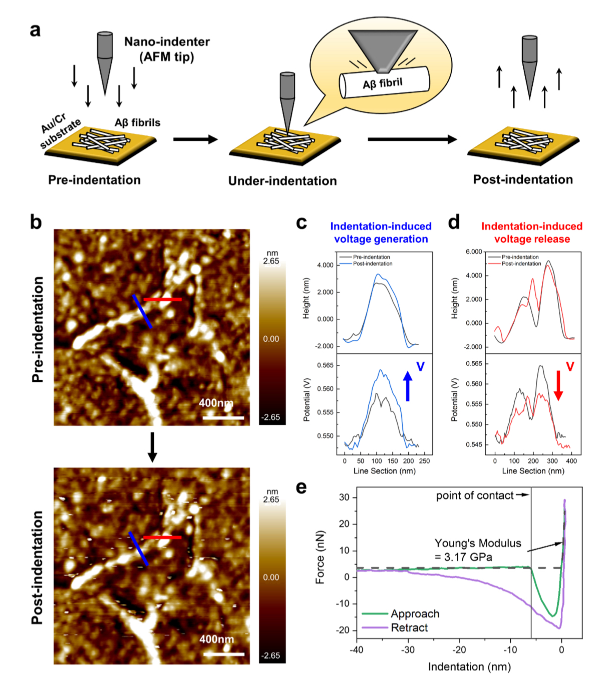
Figure S10 from „Lateral Piezoelectricity of Alzmeiner‘s Aβ Aggregates by Jinhyeong Jang et al.:
Nanoindentation test result of Aβ fibrils. Direct piezoelectric effects are tested by measuring the surface potential change before and after nano-indentation of Aβ fibrils.
(A) Schematic illustration of the nanoindentation test for Aβ fibrils coated on an Au/Cr substrate.
(B) Topgraphy of Aβ fibrils acquired before and after applying nanoindentation. The post-indentation image shows white spots resulting from physical interactions between the AFM tip and the sample, while the approximate structure of the Aβ fibrils remains intact. Height and potential line sections obtained fom the (c) blue and (d) red lines are shown in the topgraphy of Aβ fibrils. The height remains unchanged, while electrical voltage increment and decrement were observed at the blue and red lines, respectively.
(D) During the nanoindentation the Young‘s modulus of Aβ – fibrils was measured to be 3.17 GPa, closely approxmating the literature (Nanoscale 4, 4426-4429 (2012). Both KPFM and nanoindentation measurements were performed using a chemically inert conductive diamond AFM tip (CDT-FMR, NanoWorld, Switzerland). Collected force curves were analyzed using the Hertz model to estimate the elastic moduli.

*Jinhyeong Jang, Soyun Joo, Jiwon Yeom, Yonghan Jo, Jingshu Zhang, Seungbum Hong and Chan Beum Park
Lateral Piezoelectricity of Alzheimer‘s Aβ Aggregates
Advanced Science, Volume 11, Issue 39, October 024, 2406678
DOI: https://doi.org/10.1002/advs.202406678

Open Access The article “Lateral Piezoelectricity of Alzheimer‘s Aβ Aggregates” byJinhyeong Jang, Soyun Joo, Jiwon Yeom, Yonghan Jo, Jingshu Zhang, Seungbum Hong and Chan Beum Park is licensed under a Creative Commons Attribution 4.0 International License, which permits use, sharing, adaptation, distribution and reproduction in any medium or format, as long as you give appropriate credit to the original author(s) and the source, provide a link to the Creative Commons license, and indicate if changes were made. The images or other third party material in this article are included in the article’s Creative Commons license, unless indicated otherwise in a credit line to the material. If material is not included in the article’s Creative Commons license and your intended use is not permitted by statutory regulation or exceeds the permitted use, you will need to obtain permission directly from the copyright holder. To view a copy of this license, visit http://creativecommons.org/licenses/by/4.0/.

Spherulite-like microstructure observed for spin-cast P(VDF-TrFE) thin films and their ferroelectric characteristics

Ferroelectric polymer thin films crystallize in different phases and microstructures depending on fabrication conditions such as annealing temperature or layer deposition technique.*

In the article “Spherulite-like microstructure observed for spin-cast P(VDF-TrFE) thin films and their ferroelectric characteristics” Davide Disnan, Jonas Hafner, Michael Schneider and Ulrich Schmid demonstrate how the morphology of spin-cast poly(vinylidene fluoride-trifluoroethylene) (P(VDF-TrFE)) thin films changes as a function of annealing temperature from rice-like to spherulite-like microstructure, whereas the latter morphology is closer to the crystallization characteristic of poly(vinylidene-fluoride) (PVDF).*

Thin films of PVDF and P(VDF-TrFE) were analyzed at the nanometre-scale using atomic force microscopy. *

NanoWorld Pyrex-Nitride PNP-TR AFM probes were used for the ferroelectric characterization of the polymer thin films by atomic force microscopy. *

The displacement of the metal-ferroelectric-metal structure in response to the electric field applied was measured at one single point in the centre of the capacitor. For that purpose, the AFM cantilever (NanoWorld PNP-TR with a spring constant of k = 0.32 N/m made of silicon nitride (non-conductive cantilever for avoiding electrical interference caused by ground loops) was used.*

The deflection during the electrical stimulation was calibrated through the measurement of the inverse optical lever sensitivity of the probe. In order to avoid significant indentation effects, the silicon wafer surface surrounding the capacitor structure was used to land the cantilever for the calibration. *

Fig. 1 from «Spherulite-like microstructure observed for spin-cast P(VDF-TrFE) thin films and their ferroelectric characteristics» by D. Disnan et al: Spherulite-like microstructure of P(VDF-TrFE) and spherulite microstructure of PVDF. a, b Optical micrograph and AFM height image of the spherulite-like microstructure of P(VDF-TrFE). Features like needle-shaped crystals (NSC), nucleation centres (NC) and grain boundaries (GB) are highlighted on the spherulite-like microstructure surface. C ,d Analog for the spherulite microstructure of PVDF. NanoWorld Pyrex-Nitride AFM probes of the PNP-TR type were used for the Ferroelectric characterization of the polymer thin films.
Fig. 1 from «Spherulite-like microstructure observed for spin-cast P(VDF-TrFE) thin films and their ferroelectric characteristics» by D. Disnan et al:
Spherulite-like microstructure of P(VDF-TrFE) and spherulite microstructure of PVDF. a, b Optical micrograph and AFM height image of the spherulite-like microstructure of P(VDF-TrFE). Features like needle-shaped crystals (NSC), nucleation centres (NC) and grain boundaries (GB) are highlighted on the spherulite-like microstructure surface. C ,d Analog for the spherulite microstructure of PVDF.

Please follow the external link to read the full article:

*Davide Disnan, Jonas Hafner, Michael Schneider and Ulrich Schmid
Spherulite-like microstructure observed for spin-cast P(VDF-TrFE) thin films and their ferroelectric characteristics
Polymer, Volume 272, 17 April 2023, 125840
DOI: https://doi.org/10.1016/j.polymer.2023.125840

The article “Spherulite-like microstructure observed for spin-cast P(VDF-TrFE) thin films and their ferroelectric characteristics” by Davide Disnan, Jonas Hafner, Michael Schneider and Ulrich Schmid is licensed under a Creative Commons Attribution 4.0 International License, which permits use, sharing, adaptation, distribution and reproduction in any medium or format, as long as you give appropriate credit to the original author(s) and the source, provide a link to the Creative Commons license, and indicate if changes were made. The images or other third party material in this article are included in the article’s Creative Commons license, unless indicated otherwise in a credit line to the material. If material is not included in the article’s Creative Commons license and your intended use is not permitted by statutory regulation or exceeds the permitted use, you will need to obtain permission directly from the copyright holder. To view a copy of this license, visit https://creativecommons.org/licenses/by/4.0/.


Piezoelectricity of green carp scales

Today is Children’s Day in Japan and many mulit-colored carp-shaped koinobori streamers are fluttering in the wind.

So it is the perfect day to repost and share the publication “Piezoelectricity of green carp scales” by Y. Jiang et al. with you.

Piezoelectricity takes part in multiple important functions and processes in biomaterials often vital to the survival of organisms. In their publication , “Piezoelectricity of green carp scales” Y. Jiang et al. investigate the piezoelectric properties of fish scales of green carp by directly examining their morphology at nanometer levels. From the clear distinctions between the composition of the inner and outer surfaces of the scales that could be found, the authors identified the piezoelectricity to originate from the presence of hydroxyapatite which only exists on the surface of the fish scales.*

koinobori - carp streamers on children's day in Matsumoto Japan
koinobori – carp streamers on children’s day in Matsumoto Japan

These findings reveal a different mechanism of how green carp are sensitive to their surroundings and should be helpful to studies related to the electromechanical properties of marine life and the development of bio-inspired materials. As easily accessible natural polymers, fish scales can be employed as highly sensitive piezoelectric materials in high sensitive and high speed devices as well as be exploited for invasive diagnostics and other biomedical implications.*

For the harmonic responses of both 1st order and 2nd order described in this publication, NanoWorld Arrow-CONTPt AFM probes were used.

FIG. 6 from “Piezoelectricity of green carp scales “ by H. Y. Jiang et al.: First and second harmonic responses of (a) domain I and (b) domain IV. The straight line fitting for the amplitude of first harmonic response of (c) domain I and (d) domain IV by applying a series of bias. NanoWorld Arrow-CONTPt AFM probes were used.
FIG. 6 from “Piezoelectricity of green carp scales “ by H. Y. Jiang et al.: First and second harmonic responses of (a) domain I and (b) domain IV. The straight line fitting for the amplitude of first harmonic response of (c) domain I and (d) domain IV by applying a series of bias.

*Y. Jiang, F. Yen, C. W. Huang, R. B. Mei, and L. Chen
Piezoelectricity of green carp scales
AIP Advances 7, 045215 (2017)
DOI: https://doi.org/10.1063/1.4979503

Please follow this external link to access the full article: https://aip.scitation.org/doi/full/10.1063/1.4979503

Open Access The article “Piezoelectricity of green carp scales” by Y. Jiang, F. Yen, C. W. Huang, R. B. Mei and L. Chen is licensed under a Creative Commons Attribution 4.0 International License, which permits use, sharing, adaptation, distribution and reproduction in any medium or format, as long as you give appropriate credit to the original author(s) and the source, provide a link to the Creative Commons license, and indicate if changes were made. The images or other third party material in this article are included in the article’s Creative Commons license, unless indicated otherwise in a credit line to the material. If material is not included in the article’s Creative Commons license and your intended use is not permitted by statutory regulation or exceeds the permitted use, you will need to obtain permission directly from the copyright holder. To view a copy of this license, visit http://creativecommons.org/licenses/by/4.0/.