Electrically and mechanically driven rotation of polar spirals in a relaxor ferroelectric polymer

Flux-closure structures, vortices/antivortices, skyrmions, and merons in oxides, metals and polymers represent non-trivial topologies in which a local polar/magnetic order undergoes quasi-continuous spatial variations in a host crystal lattice. These structures are now extensively studied due to emergent functionalities, but the application of electrical/mechanical fields has so far only served to destroy the polar topologies of interest. *

Topology created by quasi-continuous spatial variations of a local polarization direction represents an exotic state of matter, but field-driven manipulation has been hitherto limited to creation and destruction. *In the article “Electrically and mechanically driven rotation of polar spirals in a relaxor ferroelectric polymer” Mengfan Guo, Erxiang Xu, Houbing Huang, Changqing Guo, Hetian Chen, Shulin Chen, Shan He, Le Zhou, Jing Ma, Zhonghui Shen, Ben Xu, Di Yi, Peng Gao, Ce-Wen Nan, Neil. D. Mathur and Yang Shen report that relatively small electric or mechanical fields can drive the non-volatile rotation of polar spirals in discretized microregions of the relaxor ferroelectric polymer poly(vinylidene fluoride-ran-trifluoroethylene).*

These polar spirals arise from the asymmetric Coulomb interaction between vertically aligned helical polymer chains, and can be rotated in-plane through various angles with robust retention. *

Given also that their manipulation of topological order can be detected via infrared absorption, Mengfan Guo et al.’s work suggests a new direction for the application of complex materials. *

Mengfan Guo et al. produced a 100-nm-thick monolayer of face-on lamellae with vertically aligned polymer chains by melt-recrystallizing spin-coated thin films of P(VDF-TrFE).

The resulting melt-recrystallized thin film of the relaxor ferroelectric polymer was characterized by the authors using a commercial atomic force microscope for in-plane piezo-response force microscopy (IP-PFM).

NanoWorld Platinum Iridium coated Arrow-CONTPt AFM probes (typical resonant frequency: 14 kHz, typical force constant: 0.2 N/m, typical AFM tip radius 25 nm) were used for the in-plane (IP) PFM tests and the PFM lithography tests.

For piezo-response force microscopy (PFM) imaging, Vector mode was used where AFM tips were modulated at around 240 kHz for IP imaging, with the AC voltage set at 2 V. The images obtained by Vector Mode were double checked by using dual AC resonance tracking (DART) mode and the patterns could be reproduced. *

For angle-resolved IP-PFM tests, the rotation of sample was controlled by a protractor. To ensure identical position was imaged after rotating the sample, the authors made cross-scratches as a mark on the sample surface in advance. This method was applied to locate the scanning position in other situations if Mengfan Guo et al. had to move the sample in between the scanning probe microscopic studies.

For electric-field-induced manipulations using PFM lithography, the DC voltage on AFM tip was previously edited in the software. The scan speed was set at 1.95 Hz and no AC voltage was applied during the scanning. The DC voltage was divided by film thickness (100 nm) to obtain the electric field value. And an electric field with downward direction is defined with a positive sign.

For stress-induced manipulations, the deflection value of the PFM cantilever, which is a signal from photodetector, was preset to control the stress/force applied onto the sample. The difference in deflection value between a pressed AFM cantilever and a free AFM cantilever reflects how hard the AFM tip and sample surface are pressed to each other.*

To obtain the force value F, Mengfan Guo et al. first calibrated the AFM tips by the thermal noise method, and obtain the inverse optical lever sensitivity (InvOLS) and the spring constant k of the AFM tips.

The authors also conducted a polarization analysis based on their PFM measurements. *

To obtain the nominal toroidal order evaluated by the local curvature, the obtained IP-PFM amplitude image was firstly divided into 33 × 33 arrays, and each region was then subjected to a recognition of potential domain walls and measurement of an averaged curvature radius. *

To obtain polarization maps, angle-resolved IP-PFM images were first aligned to correct spatial distortion in nanoscale measurement. Positions with specific morphological characteristics were selected as reference points to determine the coordinate. After the correction, improved angle-resolved IP-PFM phase images would be divided into 64 × 64 arrays for deriving polarization maps. *
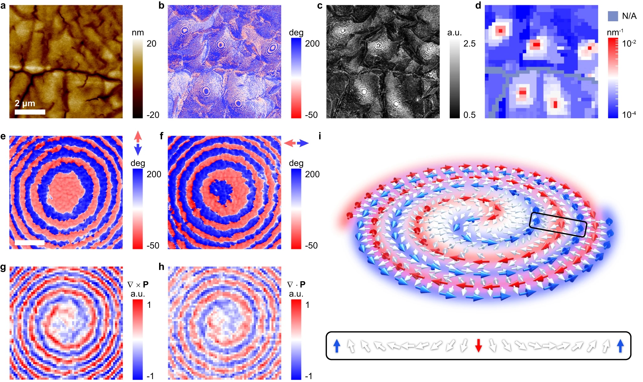
Fig. 1 from Mengfan Guo et al. (2024) “Electrically and mechanically driven rotation of polar spirals in a relaxor ferroelectric polymer”:Observation of a microregion containing an in-plane polar spiral. a Morphology of a melt-recrystallized thin film of the relaxor ferroelectric polymer. The scale bar is 2 μm. IP-PFM phase (b) and amplitude (c) images of the same area in a exhibiting concentric ring-shaped domains in curly stripe domains. d Distribution of domain wall curvatures in the same area in a–c evidencing nominal toroidal order. It is assumed that the local polarization is parallel to the nearest domain wall so that larger curvature (denoted red) reflects stronger toroidal order. IP-PFM phase images of identical concentric ring-shaped domains with the axis along vertical (e) and horizontal (f) measurement directions. The scale bar is 0.3 μm. The curl (g) and the divergence (h) of local polarization in the same area as e and f, revealing the polar spiral topology. i Schematic stereoscopic view of a CCW polar spiral, arrows represent regions of polarization. The red/blue arrows denote the polar source/sink that spirals in/out. The white arrows represent Néel rotation along the radial direction, as shown in more detail via the inset. NanoWorld Platinum Iridium coated Arrow-CONTPt AFM probes (typical resonant frequency: 14 kHz, typical force constant: 0.2 N/m, typical AFM tip radius 25 nm) were used for the in-plane (IP) PFM tests and the PFM lithography tests.
Fig. 1 from Mengfan Guo et al. (2024) “Electrically and mechanically driven rotation of polar spirals in a relaxor ferroelectric polymer”:
Observation of a microregion containing an in-plane polar spiral.
a Morphology of a melt-recrystallized thin film of the relaxor ferroelectric polymer. The scale bar is 2 μm. IP-PFM phase (b) and amplitude (c) images of the same area in a exhibiting concentric ring-shaped domains in curly stripe domains. d Distribution of domain wall curvatures in the same area in a–c evidencing nominal toroidal order. It is assumed that the local polarization is parallel to the nearest domain wall so that larger curvature (denoted red) reflects stronger toroidal order. IP-PFM phase images of identical concentric ring-shaped domains with the axis along vertical (e) and horizontal (f) measurement directions. The scale bar is 0.3 μm. The curl (g) and the divergence (h) of local polarization in the same area as e and f, revealing the polar spiral topology. i Schematic stereoscopic view of a CCW polar spiral, arrows represent regions of polarization. The red/blue arrows denote the polar source/sink that spirals in/out. The white arrows represent Néel rotation along the radial direction, as shown in more detail via the inset.

*Mengfan Guo, Erxiang Xu, Houbing Huang, Changqing Guo, Hetian Chen, Shulin Chen, Shan He, Le Zhou, Jing Ma, Zhonghui Shen, Ben Xu, Di Yi, Peng Gao, Ce-Wen Nan, Neil. D. Mathur and Yang Shen
Electrically and mechanically driven rotation of polar spirals in a relaxor ferroelectric polymer
Nature Communications volume 15, Article number: 348 (2024)
DOI: https://doi.org/10.1038/s41467-023-44395-5

The article “Electrically and mechanically driven rotation of polar spirals in a relaxor ferroelectric polymer” by Mengfan Guo, Erxiang Xu, Houbing Huang, Changqing Guo, Hetian Chen, Shulin Chen, Shan He, Le Zhou, Jing Ma, Zhonghui Shen, Ben Xu, Di Yi, Peng Gao, Ce-Wen Nan, Neil. D. Mathur and Yang Shen is licensed under a Creative Commons Attribution 4.0 International License, which permits use, sharing, adaptation, distribution and reproduction in any medium or format, as long as you give appropriate credit to the original author(s) and the source, provide a link to the Creative Commons license, and indicate if changes were made. The images or other third-party material in this article are included in the article’s Creative Commons license, unless indicated otherwise in a credit line to the material. If material is not included in the article’s Creative Commons license and your intended use is not permitted by statutory regulation or exceeds the permitted use, you will need to obtain permission directly from the copyright holder. To view a copy of this license, visit https://creativecommons.org/licenses/by/4.0/.

Quasi-one-dimensional metallic conduction channels in exotic ferroelectric topological defects

Topological objects and defects (e.g. skyrmions, domain walls, vortices,) in condensed matters have attracted a lot of interest as a field for exploring emerging exotic phenomena and functionalities.*

In materials with ferroic order, these topological objects can also be manipulated and controlled by external fields without disrupting their host lattice, making them promising elemental building blocks for potential configurable topological nanoelectronics. *

Ferroelectric topological objects provide a promising area for investigating emerging physical properties that could potentially be utilized in future nanoelectronic devices. *

In the article “Quasi-one-dimensional metallic conduction channels in exotic ferroelectric topological defects” Wenda Yang, Guo Tian, Yang Zhang, Fei Xue, Dongfeng Zheng, Luyong Zhang, Yadong Wang, Chao Chen, Zhen Fan, Zhipeng Hou, Deyang Chen, Jinwei Gao, Min Zeng, Minghui Qin, Long-Qing Chen, Xingsen Gao and Jun-Ming Liu demonstrate the existence of metallic conduction superfine (<3 nm) channels in two types of exotic topological defects, namely a quadrant vortex core or simply vortex core and a quadrant center domain core or simply center core, in an array of BiFeO3 (BFO) nanoislands.*

The authors discover via the phase-field simulation that the superfine metallic conduction channels along the center cores arise from the screening charge carriers confined at the core region, whereas the high conductance of vortex cores results from a field-induced twisted state. These conducting channels can be reversibly created and deleted by manipulating the two topological states via electric field, leading to an apparent electroresistance effect with an on/off ratio higher than 103.*

The findings by Wenda Yang et al. open up the possibility of using these functional one-dimensional topological objects in high-density nanoelectronic devices, e.g. nonvolatile memory.*

NanoWorld PlatinumIdridium5 coated Arrow-EFM AFM probes were used to examine the domain structures by vector piezoresponse force microscopy (PFM). By using vector PFM mode, the authors could simultaneously map the vertical and lateral piezoresponse signals from the nanoisland one by one.*

NanoWorld Conductive Diamond coated AFM probes CDT-NCHR were used for the conductive current distribution maps, current–voltage (I–V) measurements that were characterized by conductive atomic force microscopy (C-AFM).

Fig. 2 from “Quasi-one-dimensional metallic conduction channels in exotic ferroelectric topological defects” by Wenda Yang et al.:
The domain structures and corresponding conductive properties for both a vortex and a center topological states confined in two nanoislands.
a, b PFM and C-AFM images for both a vortex state (a) and a center state (b), the micrographs from the left to the right are PFM vertical phase images illustrating the uniform upward vertical polarization components for both nanoislands, the PFM lateral phase images recorded at sample rotation of 0o and 90o to evaluate the directions of lateral polarization components respectively along x axis ([100] axis) and y axis ([100] axis), the lateral polarization vector direction maps derived from the PFM data, and corresponding C-AFM maps. The thick arrows aside the PFM images mark the directions of the cantilever for each PFM scan, and the fine arrows inside the images mark the directions of polarization components perpendicular to the directions of the cantilever. c, d Extracted current spatial profiles from the C-AFM maps for both the vortex (c) and the center (d) cores, extracted from a and b, respectively. The inserts in c and d illustrate the C-AFM maps and schematic local polarization configurations for the two topological cores. e Temperature-dependent conductive current (I–V) curves for both topological cores and domain walls.*

*Wenda Yang, Guo Tian, Yang Zhang, Fei Xue, Dongfeng Zheng, Luyong Zhang, Yadong Wang, Chao Chen, Zhen Fan, Zhipeng Hou, Deyang Chen, Jinwei Gao, Min Zeng, Minghui Qin, Long-Qing Chen, Xingsen Gao and Jun-Ming Liu
Quasi-one-dimensional metallic conduction channels in exotic ferroelectric topological defects
Nature Communications volume 12, Article number: 1306 (2021)
DOI: https://doi.org/10.1038/s41467-021-21521-9

Please follow this external link to read the full article: https://rdcu.be/cg0JY

Open Access : The article “Quasi-one-dimensional metallic conduction channels in exotic ferroelectric topological defects” by Wenda Yang, Guo Tian, Yang Zhang, Fei Xue, Dongfeng Zheng, Luyong Zhang, Yadong Wang, Chao Chen, Zhen Fan, Zhipeng Hou, Deyang Chen, Jinwei Gao, Min Zeng, Minghui Qin, Long-Qing Chen, Xingsen Gao and Jun-Ming Liu is licensed under a Creative Commons Attribution 4.0 International License, which permits use, sharing, adaptation, distribution and reproduction in any medium or format, as long as you give appropriate credit to the original author(s) and the source, provide a link to the Creative Commons license, and indicate if changes were made. The images or other third party material in this article are included in the article’s Creative Commons license, unless indicated otherwise in a credit line to the material. If material is not included in the article’s Creative Commons license and your intended use is not permitted by statutory regulation or exceeds the permitted use, you will need to obtain permission directly from the copyright holder. To view a copy of this license, visit https://creativecommons.org/licenses/by/4.0/.

Influence of orientation and ferroelectric domains on the photochemical reactivity of La2Ti2O7

In the article “Influence of orientation and ferroelectric domains on the photochemical reactivity of La2Ti2O7” Mingyi Zhang, Paul A. Salvador and Gregory S. Rohrer describe how they measured the effects of crystal orientation and ferroelectric domain structure on the photochemical reactivity of La2Ti2O7. *

The reactivity is greatest on (001) surfaces (this is the orientation of the layers in this (110)p layered perovskite structure) while surfaces perpendicular to this orientation have the least reactivity. Complex domain structures were observed within the grains, but they appeared to have no effect on the photocathodic reduction of silver, in contrast to previous observations on other ferroelectrics. La2Ti2O7 is an example of a ferroelectric oxide in which the crystal orientation has a greater influence on the photochemical reactivity than polarization from the internal domain structure. *

NanoWorld™ conductive Platinum Iridium coated Arrow-EFM AFM probes were used for the Piezo-force microscopy (PFM) that was used to determine the ferroelectric domain structure on the surface. *

The ferroelectric domains on the surface were found to have irregular shapes and there was no correlation between the pattern of silver reduction and the domain shape. The results indicate that the ferroelectric polarization of La2Ti2O7 does not alter the reactivity enough to overcome the influence of the anisotropic crystal structure. *

Fig. 6 a and b from “Influence of orientation and ferroelectric domains on the photochemical reactivity of La2Ti2O7” by Mingyi Zhang et al.
A La2Ti2O7 grain imaged with different modalities. (a) a PFM out-of-plane amplitude image. (b) a PFM out-of-plane phase image. A meandering black line in (a), marked by the arrow, corresponds to a change from light to dark contrast in the phase image. The dark (light) contrast corresponds to regions with -180° (0°) phase shift.  NanoWorld conductive Arrow-EFM AFM probes were used for the piezo-force microscopy.

Please have a look at the full article cited below for the full figure
Fig. 6 a and b from “Influence of orientation and ferroelectric domains on the photochemical reactivity of La2Ti2O7” by Mingyi Zhang et al.
A La2Ti2O7 grain imaged with different modalities. (a) a PFM out-of-plane amplitude image. (b) a PFM out-of-plane phase image. A meandering black line in (a), marked by the arrow, corresponds to a change from light to dark contrast in the phase image. The dark (light) contrast corresponds to regions with -180° (0°) phase shift. Please have a look at the full article cited below for the full figure

*Mingyi Zhang, Paul A. Salvador and Gregory S.Rohrer
Influence of orientation and ferroelectric domains on the photochemical reactivity of La2Ti2O7
Journal of the European Ceramic Society (2020)
DOI: https://doi.org/10.1016/j.jeurceramsoc.2020.09.020

Please follow this external link to read the full article https://www.sciencedirect.com/science/article/pii/S0955221920307445

Open Access : The article “Influence of orientation and ferroelectric domains on the photochemical reactivity of La2Ti2O7” by Mingyi Zhang, Paul A. Salvador, Gregory S. Rohrer is licensed under a Creative Commons Attribution 4.0 International License, which permits use, sharing, adaptation, distribution and reproduction in any medium or format, as long as you give appropriate credit to the original author(s) and the source, provide a link to the Creative Commons license, and indicate if changes were made. The images or other third party material in this article are included in the article’s Creative Commons license, unless indicated otherwise in a credit line to the material. If material is not included in the article’s Creative Commons license and your intended use is not permitted by statutory regulation or exceeds the permitted use, you will need to obtain permission directly from the copyright holder. To view a copy of this license, visit https://creativecommons.org/licenses/by/4.0/.