Flexible Polyurethane Foams Modified with Novel Coconut Monoglycerides-Based Polyester Polyols

The products of the polyurethane (PU) industry such as foams, coatings and adhesives are numerous and can be found in many areas of everyday life. *

Polyols are an essential component in the production of polyurethane. Nowadays they mostly come from petroleum products. *

In view of potential risk factors such as the running out of fossil fuels, supply chain issues, environmental concerns and economic risks it is important to develop alternatives as substitutes and supplements to the existing petroleum derived polyols. *

Vegetable oils can be used to manufacture biobased polyols and various oils such as linseed oil, rapeseed oil, canola oil, grapeseed oil, corn oil, rice bran oil, palm oil, olive oil, castor oil and soybean oil have already been used to make polyols for different purposes.*

Most of the polyols derived from vegetable oils that are already commercially available are made from soybean and castor oil and are mainly used for rigid PU foam applications. *

So far biobased materials for flexible polyurethane foams (FPUFs) have not been studied as much as their rigid counterpart. This is because, due to their chemical composition, there are limits to how much biobased materials can be used in the flexible foam without having an undesired effect on the foam’s mechanical properties. *

Coconut oil is not often used to manufacture flexible foam because the coconut oil’s high level of saturation makes it less compatible with many common methods of creating polyols, as the widely used polyol-forming processes mostly rely on the unsaturation of vegetable oil for functionalization.

To make coconut monoglycerides (CMG) or other plant-based oils usable for polyol-forming processes they need to fulfil the same structural requirements as the fossil-based products. *

In the article “Flexible Polyurethane Foams Modified with Novel Coconut Monoglycerides-Based Polyester Polyols “ Christine Joy M. Omisol, Blessy Joy M. Aguinid, Gerson Y. Abilay, Dan Michael Asequia, Tomas Ralph Tomon, Karyl Xyrra Sabulbero, Daisy Jane Erjeno, Carlo Kurt Osorio, Shashwa Usop, Roberto Malaluan, Gerard Dumancas, Eleazer P. Resurreccion, Alona Lubguban, Glenn Apostol, Henry Siy, Arnold C. Alguno, and Arnold Lubguban describe how they investigated the potential of coconut monoglycerides (CMG) as a polyol raw material specifically for flexible polyurethane foam (FPUF) applications.*

The authors synthesized high-molecular-weight polyester polyols from coconut monoglycerides (CMG), a coproduct of fatty acid production from coconut oil, via polycondensation at different mass ratios of CMG with 1:5 glycerol:phthalic anhydride.*

The resulting CMG-based polyols were shown to work well in making flexible foam. *

Fourier transform infrared (FTIR) spectroscopy and atomic force microscopy (AFM) were used for the foam characterization. *

The modification of the foam formulation increased the monodentate and bidentate urea groups, shown using Fourier transform infrared (FTIR) spectroscopy, that promoted microphase separation in the foam matrix, confirmed using atomic force microscopy (AFM) and differential scanning calorimetry (DSC). *

Atomic force microscopy (AFM) was used to evaluate the hard–soft domains phase separation of the foam. *

The atomic force microscope was operated at a scan rate of 1.0 Hz in non-contact mode using NanoWorld Pointprobe® NCHR Silicon AFM probes for standard tapping mode applications. (typical resonance frequency 320 kHz, typical force constant 42 N/m). *

It could be shown that density of the CMGPOL-modified polyurethane foams (CPFs) decreased, while a significant improvement in their tensile and compressive properties was observed. *

The investigations by Christine Joy M. Omisol et al. resulted in a new sustainable polyol raw material that can be used to modify petroleum-based foam and produce flexible foams with varying properties that can be tailored to meet specific requirements. *
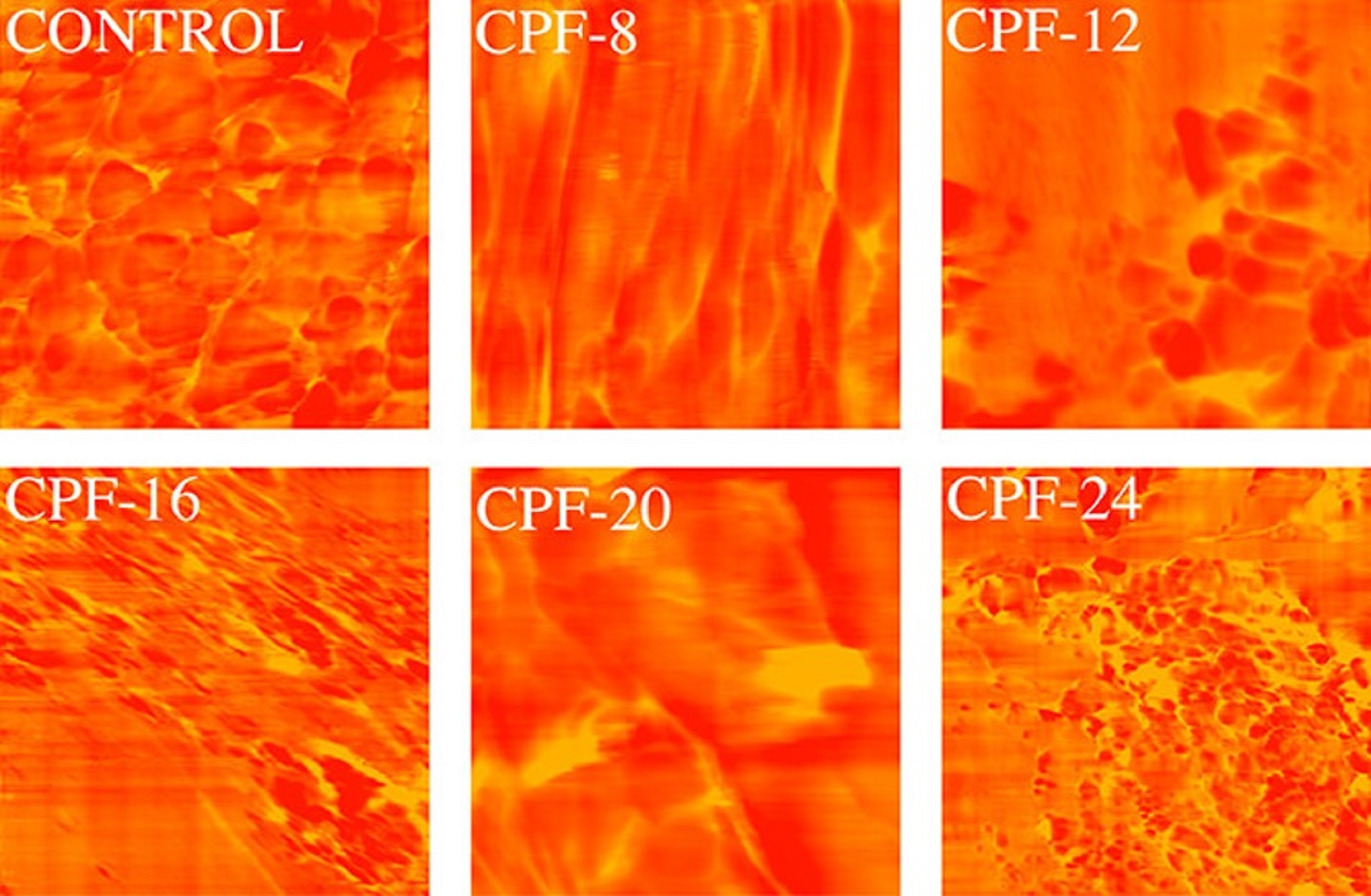
Figure 11 from Christine Joy M. Omisol et al (2024) “Flexible Polyurethane Foams Modified with Novel Coconut Monoglycerides-Based Polyester Polyols”:Atomic force microscopy (AFM) phase images of CMGPOL-modified polyurethane foams (CPF) and control foam measured with a size scan of 3 μm × 3 μm showing soft and hard regions represented by red and yellow colors, respectively. The foam samples in Figure 11 that exhibited a relatively high degree of microphase separation compared with other samples are CPF-8 and CPF-20. These foams appear to have relatively lighter areas of urea-rich regions separated more prominently from the darker, polyol-rich regions. In contrast, the control foam and CPF-16 show more dispersed hard and soft domains. CPF-24 and CPF-12 are at the middle of the scale, displaying light regions but with more dispersion than CPF-8 and CPF-20. These observations from the phase images of the foam samples are in agreement with the monodentate and bidentate urea contents of the samples, wherein the foams that exhibit greater H-bonding also manifest a higher degree of microphase separation. The same results were obtained by Baghban et al. NanoWorld Pointprobe® NCHR standard tapping mode/non-contact mode silicon AFM probes were used for the foam characterizations with atomic force microscopy.
Figure 11 from Christine Joy M. Omisol et al (2024) “Flexible Polyurethane Foams Modified with Novel Coconut Monoglycerides-Based Polyester Polyols”:
Atomic force microscopy (AFM) phase images of CMGPOL-modified polyurethane foams (CPF) and control foam measured with a size scan of 3 μm × 3 μm showing soft and hard regions represented by red and yellow colors, respectively.

*Christine Joy M. Omisol, Blessy Joy M. Aguinid, Gerson Y. Abilay, Dan Michael Asequia, Tomas Ralph Tomon, Karyl Xyrra Sabulbero, Daisy Jane Erjeno, Carlo Kurt Osorio, Shashwa Usop, Roberto Malaluan, Gerard Dumancas, Eleazer P. Resurreccion, Alona Lubguban, Glenn Apostol, Henry Siy, Arnold C. Alguno, and Arnold Lubguban
Flexible Polyurethane Foams Modified with Novel Coconut Monoglycerides-Based Polyester Polyols
ACS Omega 2024, 9, 4, 4497–4512
DOI: https://doi.org/10.1021/acsomega.3c07312

The article “Flexible Polyurethane Foams Modified with Novel Coconut Monoglycerides-Based Polyester Polyols” by Christine Joy M. Omisol, Blessy Joy M. Aguinid, Gerson Y. Abilay, Dan Michael Asequia, Tomas Ralph Tomon, Karyl Xyrra Sabulbero, Daisy Jane Erjeno, Carlo Kurt Osorio, Shashwa Usop, Roberto Malaluan, Gerard Dumancas, Eleazer P. Resurreccion, Alona Lubguban, Glenn Apostol, Henry Siy, Arnold C. Alguno and Arnold Lubguban is licensed under a Creative Commons Attribution 4.0 International License, which permits use, sharing, adaptation, distribution and reproduction in any medium or format, as long as you give appropriate credit to the original author(s) and the source, provide a link to the Creative Commons license, and indicate if changes were made. The images or other third-party material in this article are included in the article’s Creative Commons license, unless indicated otherwise in a credit line to the material. If material is not included in the article’s Creative Commons license and your intended use is not permitted by statutory regulation or exceeds the permitted use, you will need to obtain permission directly from the copyright holder. To view a copy of this license, visit https://creativecommons.org/licenses/by/4.0/.

Electrochemically Synthesized Poly(3-hexylthiophene) Nanowires as Photosensitive Neuronal Interfaces

Poly(3-hexylthiophene) (P3HT), a hole-conducting polymer, generates a lot of interest especially because of its excellent optoelectronic properties (such as good electrical conductivity and high extinction coefficient) and good processability, which make this polymer an excellent choice for building organic optoelectronic devices (e.g., organic solar cells). *

P3HT films and nanoparticles have also been used to restore the photosensitivity of retinal neurons. *

For their article “Electrochemically Synthesized Poly(3-hexylthiophene) Nanowires as Photosensitive Neuronal Interfaces” Szilveszter Gáspár, Tiziana Ravasenga, Raluca-Elena Munteanu, Sorin David, Fabio Benfenati, and Elisabetta Colombo investigated the template-assisted electrochemical synthesis of P3HT nanowires doped with tetrabutylammonium hexafluorophosphate (TBAHFP) and their biocompatibility with primary neurons. *

They were able to show that template-assisted electrochemical synthesis can relatively easily turn 3-hexylthiophene (3HT) into longer (e.g., 17 ± 3 µm) or shorter (e.g., 1.5 ± 0.4 µm) P3HT nanowires with an average diameter of 196 ± 55 nm (determined by the used template) and that the nanowires produce measurable photocurrents following illumination. *

The fact that template-assisted electrochemical synthesis combines polymerization, doping, and polymer nanostructuring into one, relatively simple step is the most important advantage of this method. The possibility of easily tuning the length of the produced nanowires represents another important advantage. *

The authors were also able to demonstrate that primary cortical neurons can be grown onto P3HT nanowires drop-casted on a glass substrate without relevant changes in their viability and electrophysiological properties, indicating that P3HT nanowires obtained by template-assisted electrochemical synthesis represent a promising neuronal interface for photostimulation. *

Szilveszter Gáspár  et al. proved the biocompability of the obtained P3HT nanowires upon incubation for different periods with primary neuronal cultures. They demonstrated that their presence does not affect the membrane properties of the neurons or the excitability of the neurons as evaluated by patch-clamp experiments. These results show the potential of the described synthesis methodology to fabricate injectable P3HT-based photosensitive nanowires with high biocompatibility, ultimately paving the way for their exploitation for neuronal photostimulation. *

Atomic Force Microscopy (AFM) was used to characterize P3HT nanowires drop-casted onto glass coverslips. *

The Atomic Force Microscopy images were obtained in air and in intermittent contact-mode using line rates as slow as 0.2 Hz and NanoWorld Pointprobe® NCSTR silicon soft-tapping AFM probes (typical values: resonant frequency 160 kHz, force constant 7.2 N m). The ratio between the set-point amplitude and the free amplitude of the AFM cantilever was set to 0.5–0.6. The obtained AFM images were used to determine both the lengths and the diameters of the nanowires. *

Figure 3 from “Electrochemically Synthesized Poly(3-hexylthiophene) Nanowires as Photosensitive Neuronal Interfaces” by Szilveszter Gáspár et al.: AFM images of “long” P3HT nanowires (A) and of “short” P3HT nanowires (B). NanoWorld Pointprobe NCSTR soft-tapping mode probes were used.
Figure 3 from “Electrochemically Synthesized Poly(3-hexylthiophene) Nanowires as Photosensitive Neuronal Interfaces” by Szilveszter Gáspár et al.:
AFM images of “long” P3HT nanowires (A) and of “short” P3HT nanowires (B).

*Szilveszter Gáspár, Tiziana Ravasenga, Raluca-Elena Munteanu, Sorin David, Fabio Benfenati, and Elisabetta Colombo
Electrochemically Synthesized Poly(3-hexylthiophene) Nanowires as Photosensitive Neuronal Interfaces
Materials 2021, 14(16), 4761, Special Issue Advanced Designs of Materials, Devices and Techniques for Biosensing
DOI: https://doi.org/10.3390/ma14164761 (please follow this external link to read the full article.)

Open Access The article “Electrochemically Synthesized Poly(3-hexylthiophene) Nanowires as Photosensitive Neuronal Interfaces” by Szilveszter Gáspár, Tiziana Ravasenga, Raluca-Elena Munteanu, Sorin David, Fabio Benfenati, and Elisabetta Colombo is licensed under a Creative Commons Attribution 4.0 International License, which permits use, sharing, adaptation, distribution and reproduction in any medium or format, as long as you give appropriate credit to the original author(s) and the source, provide a link to the Creative Commons license, and indicate if changes were made. The images or other third party material in this article are included in the article’s Creative Commons license, unless indicated otherwise in a credit line to the material. If material is not included in the article’s Creative Commons license and your intended use is not permitted by statutory regulation or exceeds the permitted use, you will need to obtain permission directly from the copyright holder. To view a copy of this license, visit http://creativecommons.org/licenses/by/4.0/.

Optimized positioning through maximized tip visibility – Arrow AFM probes screencast passes 500 views mark

The screencast about NanoWorld Arrow Silicon AFM probes held byNanoWorld AG CEO Manfred Detterbeck has just passed the 500 views mark. Congratulations Manfred!

NanoWorld Arrow™ AFM probes are designed for easy AFM tip positioning and high resolution AFM imaging and are very popular with AFM users due to the highly symetric scans that are possible with these AFM probes because of their special tip shape. They fit to all well-known commercial SPMs (Scanning Probe Microscopes) and AFMs (Atomic Force Microscopes). The Arrow AFM probe consists of an AFM probe support chip with an AFM cantilever which has a tetrahedral AFM tip at its triangular free end.

The Arrow AFM probe is entirely made of monolithic, highly doped silicon.

The unique Arrow™ shape of the AFM cantilever with the AFM tip always placed at the very end of the AFM cantilever allows easy positioning of the AFM tip on the area of interest.
The Arrow AFM probes are available for non-contact mode, contact mode and force modulation mode imaging and are also available with a conductive platinum iridum coating. Furthermore the Arrow™ AFM probe series also includes a range of tipless AFM cantilevers and AFM cantilever arrays as well as dedicated ultra-high frequency Arrow AFM probes for high speed AFM.

To find out more about the different variations please have a look at:

https://www.nanoworld.com/arrow-afm-tips

You can also find various application examples for the Arrow AFM probes in the NanoWorld blog. For a selection of these articles just click on the “Arrow AFM probes” tag on the bottom of this blog entry.