Kinetic-controlled Crystallization of α-FAPbI3 Inducing Preferred Crystallographic Orientation Enhances Photovoltaic Performance

Physical properties of the polycrystalline materials are mostly determined by their microstructure. As the crystallization process can determine the microstructure, the nucleation, and growth can also control whether the materials will be resulted in single crystalline or polycrystalline. Along with the morphological changes, anisotropic properties of the materials can also be controlled. *

As a result, preferential orientation with advanced optoelectronic properties can enhance the photovoltaic devices’ performance. *

Although incorporation of additives is one of the most studied methods to stabilize the photoactive α-phase of formamidinium lead tri-iodide (α-FAPbI3), no studies focus on how the additives affect the crystallization kinetics. *

In the article “Kinetic-Controlled Crystallization of α-FAPbI3 Inducing Preferred Crystallographic Orientation Enhances Photovoltaic Performance” along with the role of methylammonium chloride (MACl) as a “stabilizer” in the formation of α-FAPbI3, Sooeun Shin, Seongrok Seo, Seonghwa Jeong, Anir S. Sharbirin, Jeongyong Kim, Hyungju Ahn, Nam-Gyu Park and Hyunjung Shin point out the additional role as a “controller” in the crystallization kinetics. *

With microscopic observations, for example, electron backscatter diffraction and selected area electron diffraction, it is examined that higher concentration of MACl induces slower crystallization kinetics, resulting in larger grain size and [100] preferred orientation. *

Optoelectronic properties of [100] preferentially oriented grains with less non-radiative recombination, a longer lifetime of charge carriers, and lower photocurrent deviations in between each grain induce higher short-circuit current density (Jsc) and fill factor. *

Resulting MACl40 mol% attains the highest power conversion efficiency (PCE) of 24.1%.

The results provide observations of a direct correlation between the crystallographic orientation and device performance as it highlights the importance of crystallization kinetics resulting in desirable microstructures for device engineering. *

The electrical characterizations with atomic force microscopy (AFM) and conductive atomic force microscopy ( C-AFM) were done to measure the local conductance of FAPbI3 films. All measurements were performed under illumination (green LED) with a 1.3 V bias using a Pt-coated C-AFM probe (NanoWorld PlatinumIridium coated Pointprobe® CONTPt ). FTO was used for the conductive substrates. *

Conductive atomic force microscopy (C-AFM) indicated much more homogeneous photocurrent generation along the surface of (100) preferentially oriented layers. *
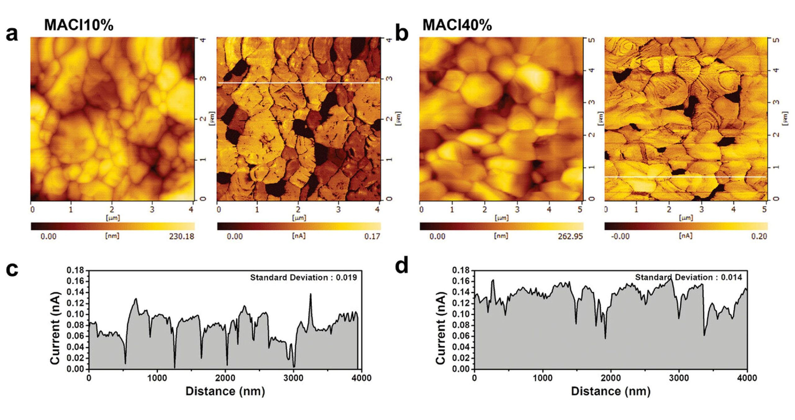
Figure 4 from Sooeun Shin et al. 2023 “Kinetic-Controlled Crystallization of α-FAPbI3 Inducing Preferred Crystallographic Orientation Enhances Photovoltaic Performance”:Electrical properties of (100)-oriented α-FAPbI3 thin films. a,b) Surface topography and photocurrent measurement by C-AFM of MACl10% and MACl40%. Measurements were taken under illumination (green LED) with a 1.3 V bias. Current images indicate that photocurrents were induced grain by grain, as each grain showed a distinct photocurrent. The photocurrent deviation for each grain is larger in MACl10% than in MACl40%, which shows a similar photocurrent grain by grain. c,d) Current line profile extracted from the current image (a and b). The standard deviation calculated from the magnitude of the photocurrent in each grain is displayed in the inset (0.019 and 0.014 nA for MACl10% and MACl40%, respectively). A low photocurrent was exhibited at the grain boundaries in both MACl10% and MACl40%. The dark areas measured within the current images are most likely distributed between grain edges. This may be caused by the formation of PbI2 or the loss of contact between the grain and the conducting substrate. NanoWorld PtIr5 coated Pointprobe® CONTPt AFM probes were used for the Electrical Characterization with conductive Atomic Force Microscopy (conductive-AFM, C-AFM) and for the atomic force microscopy.
Figure 4 from Sooeun Shin et al. 2023 “Kinetic-Controlled Crystallization of α-FAPbI3 Inducing Preferred Crystallographic Orientation Enhances Photovoltaic Performance”:
Electrical properties of (100)-oriented α-FAPbI3 thin films. a,b) Surface topography and photocurrent measurement by C-AFM of MACl10% and MACl40%. Measurements were taken under illumination (green LED) with a 1.3 V bias. Current images indicate that photocurrents were induced grain by grain, as each grain showed a distinct photocurrent. The photocurrent deviation for each grain is larger in MACl10% than in MACl40%, which shows a similar photocurrent grain by grain. c,d) Current line profile extracted from the current image (a and b). The standard deviation calculated from the magnitude of the photocurrent in each grain is displayed in the inset (0.019 and 0.014 nA for MACl10% and MACl40%, respectively). A low photocurrent was exhibited at the grain boundaries in both MACl10% and MACl40%. The dark areas measured within the current images are most likely distributed between grain edges. This may be caused by the formation of PbI2 or the loss of contact between the grain and the conducting substrate.

*Sooeun Shin, Seongrok Seo, Seonghwa Jeong, Anir S. Sharbirin, Jeongyong Kim, Hyungju Ahn, Nam-Gyu Park and Hyunjung Shin
Kinetic-Controlled Crystallization of α-FAPbI3 Inducing Preferred Crystallographic Orientation Enhances Photovoltaic Performance
Advanced Science, Volume 10, Issue 14, May 17, 2023, 2300798
DOI: https://doi.org/10.1002/advs.202300798

Open Access The article “Kinetic-Controlled Crystallization of α-FAPbI3 Inducing Preferred Crystallographic Orientation Enhances Photovoltaic Performance” Sooeun Shin, Seongrok Seo, Seonghwa Jeong, Anir S. Sharbirin, Jeongyong Kim, Hyungju Ahn, Nam-Gyu Park and Hyunjung Shin is licensed under a Creative Commons Attribution 4.0 International License, which permits use, sharing, adaptation, distribution and reproduction in any medium or format, as long as you give appropriate credit to the original author(s) and the source, provide a link to the Creative Commons license, and indicate if changes were made. The images or other third party material in this article are included in the article’s Creative Commons license, unless indicated otherwise in a credit line to the material. If material is not included in the article’s Creative Commons license and your intended use is not permitted by statutory regulation or exceeds the permitted use, you will need to obtain permission directly from the copyright holder. To view a copy of this license, visit http://creativecommons.org/licenses/by/4.0/.

Quasi-one-dimensional metallic conduction channels in exotic ferroelectric topological defects

Topological objects and defects (e.g. skyrmions, domain walls, vortices,) in condensed matters have attracted a lot of interest as a field for exploring emerging exotic phenomena and functionalities.*

In materials with ferroic order, these topological objects can also be manipulated and controlled by external fields without disrupting their host lattice, making them promising elemental building blocks for potential configurable topological nanoelectronics. *

Ferroelectric topological objects provide a promising area for investigating emerging physical properties that could potentially be utilized in future nanoelectronic devices. *

In the article “Quasi-one-dimensional metallic conduction channels in exotic ferroelectric topological defects” Wenda Yang, Guo Tian, Yang Zhang, Fei Xue, Dongfeng Zheng, Luyong Zhang, Yadong Wang, Chao Chen, Zhen Fan, Zhipeng Hou, Deyang Chen, Jinwei Gao, Min Zeng, Minghui Qin, Long-Qing Chen, Xingsen Gao and Jun-Ming Liu demonstrate the existence of metallic conduction superfine (<3 nm) channels in two types of exotic topological defects, namely a quadrant vortex core or simply vortex core and a quadrant center domain core or simply center core, in an array of BiFeO3 (BFO) nanoislands.*

The authors discover via the phase-field simulation that the superfine metallic conduction channels along the center cores arise from the screening charge carriers confined at the core region, whereas the high conductance of vortex cores results from a field-induced twisted state. These conducting channels can be reversibly created and deleted by manipulating the two topological states via electric field, leading to an apparent electroresistance effect with an on/off ratio higher than 103.*

The findings by Wenda Yang et al. open up the possibility of using these functional one-dimensional topological objects in high-density nanoelectronic devices, e.g. nonvolatile memory.*

NanoWorld PlatinumIdridium5 coated Arrow-EFM AFM probes were used to examine the domain structures by vector piezoresponse force microscopy (PFM). By using vector PFM mode, the authors could simultaneously map the vertical and lateral piezoresponse signals from the nanoisland one by one.*

NanoWorld Conductive Diamond coated AFM probes CDT-NCHR were used for the conductive current distribution maps, current–voltage (I–V) measurements that were characterized by conductive atomic force microscopy (C-AFM).

Fig. 2 from “Quasi-one-dimensional metallic conduction channels in exotic ferroelectric topological defects” by Wenda Yang et al.:
The domain structures and corresponding conductive properties for both a vortex and a center topological states confined in two nanoislands.
a, b PFM and C-AFM images for both a vortex state (a) and a center state (b), the micrographs from the left to the right are PFM vertical phase images illustrating the uniform upward vertical polarization components for both nanoislands, the PFM lateral phase images recorded at sample rotation of 0o and 90o to evaluate the directions of lateral polarization components respectively along x axis ([100] axis) and y axis ([100] axis), the lateral polarization vector direction maps derived from the PFM data, and corresponding C-AFM maps. The thick arrows aside the PFM images mark the directions of the cantilever for each PFM scan, and the fine arrows inside the images mark the directions of polarization components perpendicular to the directions of the cantilever. c, d Extracted current spatial profiles from the C-AFM maps for both the vortex (c) and the center (d) cores, extracted from a and b, respectively. The inserts in c and d illustrate the C-AFM maps and schematic local polarization configurations for the two topological cores. e Temperature-dependent conductive current (I–V) curves for both topological cores and domain walls.*

*Wenda Yang, Guo Tian, Yang Zhang, Fei Xue, Dongfeng Zheng, Luyong Zhang, Yadong Wang, Chao Chen, Zhen Fan, Zhipeng Hou, Deyang Chen, Jinwei Gao, Min Zeng, Minghui Qin, Long-Qing Chen, Xingsen Gao and Jun-Ming Liu
Quasi-one-dimensional metallic conduction channels in exotic ferroelectric topological defects
Nature Communications volume 12, Article number: 1306 (2021)
DOI: https://doi.org/10.1038/s41467-021-21521-9

Please follow this external link to read the full article: https://rdcu.be/cg0JY

Open Access : The article “Quasi-one-dimensional metallic conduction channels in exotic ferroelectric topological defects” by Wenda Yang, Guo Tian, Yang Zhang, Fei Xue, Dongfeng Zheng, Luyong Zhang, Yadong Wang, Chao Chen, Zhen Fan, Zhipeng Hou, Deyang Chen, Jinwei Gao, Min Zeng, Minghui Qin, Long-Qing Chen, Xingsen Gao and Jun-Ming Liu is licensed under a Creative Commons Attribution 4.0 International License, which permits use, sharing, adaptation, distribution and reproduction in any medium or format, as long as you give appropriate credit to the original author(s) and the source, provide a link to the Creative Commons license, and indicate if changes were made. The images or other third party material in this article are included in the article’s Creative Commons license, unless indicated otherwise in a credit line to the material. If material is not included in the article’s Creative Commons license and your intended use is not permitted by statutory regulation or exceeds the permitted use, you will need to obtain permission directly from the copyright holder. To view a copy of this license, visit https://creativecommons.org/licenses/by/4.0/.

Flexible 3D Electrodes of Free-Standing TiN Nanotube Arrays Grown by Atomic Layer Deposition with a Ti Interlayer as an Adhesion Promoter

Nanostructured electrodes and their flexible integrated systems have great potential for many applications, including electrochemical energy storage, electrocatalysis and solid-state memory devices, given their ability to improve faradaic reaction sites by large surface area. Although many processing techniques have been employed to fabricate nanostructured electrodes on to flexible substrates, these present limitations in terms of achieving flexible electrodes with high mechanical stability.*

In the study “Flexible 3D Electrodes of Free-Standing TiN Nanotube Arrays Grown by Atomic Layer Deposition with a Ti Interlayer as an Adhesion Promoter” by Seokjung Yun, Sang-Joon Kim, Jaesung Youn, Hoon Kim, Jeongjae Ryu, Changdeuck Bae, Kwangsoo No and Seungbum Hong, the adhesion, mechanical properties and flexibility of TiN nanotube arrays on a Pt substrate were improved using a Ti interlayer. Highly ordered and well aligned TiN nanotube arrays were fabricated on a Pt substrate using a template-assisted method with an anodic aluminum oxide (AAO) template and atomic layer deposition (ALD) system.*

The authors show that with the use of a Ti interlayer between the TiN nanotube arrays and Pt substrate, the TiN nanotube arrays could perfectly attach to the Pt substrate without delamination and faceted phenomena. Furthermore, the I-V curve measurements confirmed that the electric contact between the TiN nanotube arrays and substrate for use as an electrode was excellent, and its flexibility was also good for use in flexible electronic devices. Future efforts will be directed toward the fabrication of embedded electrodes in flexible plastic substrates by employing the concepts demonstrated in this study.*

The presented strategy provides a new class of nanostructured 3D electrodes to overcome critical mechanical stability, thus providing a great potential platform for application in a flexible integrated device.*

Topography and transport properties were investigated using a conductive atomic force microscope with NanoWorld Pointprobe® EFM AFM probes ( Pt-coated conductive AFM tips).*

Figure 5 from “Flexible 3D Electrodes of Free-Standing TiN Nanotube Arrays Grown by Atomic Layer Deposition with a Ti Interlayer as an Adhesion Promoter” by Seokjung Yun et al.:
Analysis of TiN NTs/ Ti / Pt samples (a) XRD, (b) schematic of C-AFM setup, (c) AFM height image, and (d) local I-V curve by C-AFM.

*Seokjung Yun, Sang-Joon Kim, Jaesung Youn, Hoon Kim, Jeongjae Ryu, Changdeuck Bae, Kwangsoo No and Seungbum Hong
Flexible 3D Electrodes of Free-Standing TiN Nanotube Arrays Grown by Atomic Layer Deposition with a Ti Interlayer as an Adhesion Promoter
Nanomaterials 2020, 10, 409
DOI: 10.3390/nano10030409

Please follow this external link for access to the full article: https://doi.org/10.3390/nano10030409

Open Access The article “Flexible 3D Electrodes of Free-Standing TiN Nanotube Arrays Grown by Atomic Layer Deposition with a Ti Interlayer as an Adhesion Promoter“ by Seokjung Yun, Sang-Joon Kim, Jaesung Youn, Hoon Kim, Jeongjae Ryu, Changdeuck Bae, Kwangsoo No and Seungbum Hong is licensed under a Creative Commons Attribution 4.0 International License, which permits use, sharing, adaptation, distribution and reproduction in any medium or format, as long as you give appropriate credit to the original author(s) and the source, provide a link to the Creative Commons license, and indicate if changes were made. The images or other third party material in this article are included in the article’s Creative Commons license, unless indicated otherwise in a credit line to the material. If material is not included in the article’s Creative Commons license and your intended use is not permitted by statutory regulation or exceeds the permitted use, you will need to obtain permission directly from the copyright holder. To view a copy of this license, visit http://creativecommons.org/licenses/by/4.0/.