Type: USC-F5-k30

Ultra-Short Cantilevers (for High-Speed AFM)

Logo
Cantilever Data Value Range*
Resonance Frequency 5000 kHz 4000 - 6000 kHz
Force Constant 30 N/m 20 - 50 N/m
Length 10 µm 9 - 11 µm
Mean Width 5 µm 4.5 - 5.5 µm
Thickness 0.68 µm 0.66 - 0.7 µm

This AFM probe has alignment grooves on the back side of the support chip.

USC cantilever 3D view

USC cantilever 3D view More images

Product Description

NanoWorld® Ultra-Short Cantilevers (USC) for High-Speed AFM (HS-AFM) combine very small AFM cantilevers capable of resonating in the MHz regime and a very sharp and wear resistant AFM tip.
 
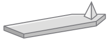
The AFM cantilever of the USC series is rectangular and made of a quartz-like material. A gold layer is deposited on both sides of the AFM cantilever in order to enhance the reflectance of the laser beam, but the AFM tip remains uncoated.

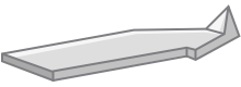
The wear resistant AFM tip has been developed together with nanotools GmbH and sustains high velocity scans over long distances. It is made of High Density Carbon/Diamond Like Carbon (HDC/DLC) material which is hard and wear resistant. It has a height of 2.5 microns and a radius of curvature smaller than 10 nm. The aspect ratio is in the order of 5 : 1 and the tilt compensation is 8° ensuring more symmetric AFM images.

The silicon support chip is of standard dimensions (1.6 mm x 3.4 mm x 0.3 mm). Additionally, it has etched and lowered corners in order to avoid contact between the support chip and the sample when scanning. Moreover it features alignment grooves on the back side of the silicon support chip which ensure replacement of the AFM probes without major adjustment of the laser beam when used in conjunction with the alignment chip.

The type USC-F5-k30 is mainly designed for High-Speed AFM applications in non-contact mode in air but can also be used for other applications.

Tip shape: Cone Shaped

Coating: Reflective Gold

Gold Reflex Coating

The gold reflex coating consists of a 30 nm thick gold layer deposited on both sides of the AFM cantilevers which enhances the reflectance of the laser beam. Furthermore it prevents light from interfering within the AFM cantilever. As the coating is almost stress-free the bending of the AFM cantilevers due to stress is less than 2 degrees

The AFM tip remains uncoated.

Order Codes

Order Code Quantity Data Sheet
USC-F5-k30-10 10 Nominal values

System limitations: Due to their small AFM cantilever sizes and their very high resonance frequencies USC probes currently cannot be used in all commercially available SPMs and AFMs. Only AFMs with a sufficiently small laser spot and electronics that are capable of dealing with high resonance frequencies of up to 5 MHz are able to work with the USC probes. If in doubt whether these AFM probes can be used in your AFM please check back with us or with your AFM manufacturer.

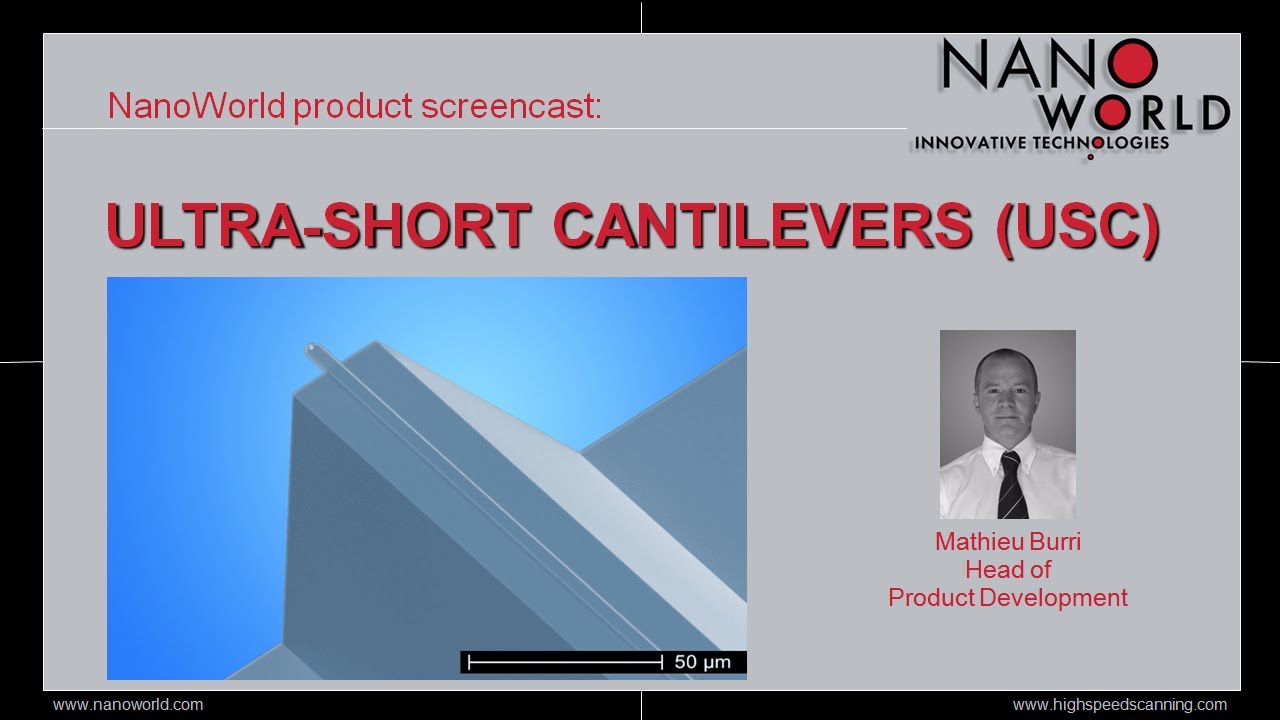
Product Screencast NanoWorld® Ultra-Short Cantilevers (USC) for High Speed Scanning

Subscribe to NanoWorld® Youtube Channel

Scientific publications mentioning use of this AFM probe


Miyata, Kazuki, Kosuke Adachi, Naoyuki Miyashita, Keisuke Miyazawa, Adam S. Foster, and Takeshi Fukuma
High-Speed Three-Dimensional Scanning Force Microscopy Visualization of Subnanoscale Hydration Structures on Dissolving Calcite Step Edges
Nano Letters. 2024 Aug 26;24(35):10842-9
DOI: https://doi.org/10.1021/acs.nanolett.4c02368


Hafner, Jonas Alexander
Ferroelectric polymer thin films for MEMS applications: towards soft and high-speed AFM probes
PhD diss., Technische Universität Wien, 2021
https://web.archive.org/web/20210501081515id_/https://repositum.tuwien.at/bitstream/20.500.12708/16943/1/Hafner%20Jonas%20Alexander%20-%202021%20-%20Ferroelectric%20Polymer%20Thin%20Films%20for%20MEMS...pdf


Konrad, Sebastian F., Willem Vanderlinden, Wout Frederickx, Tine Brouns, Björn H. Menze, Steven De Feyter, and Jan Lipfert
High-throughput AFM analysis reveals unwrapping pathways of H3 and CENP-A nucleosomes
 Nanoscale. 2021;13(10):5435-47
DOI: 10.1039/D0NR08564B


Konrad, Sebastian F
Revealing nucleosome conformations by AFM imaging and large-scale data analysis
PhD diss., lmu, 2021
https://edoc.ub.uni-muenchen.de/28949/7/Konrad_Sebastian.pdf


Hirata, Kaito, Takumi Igarashi, Keita Suzuki, Keisuke Miyazawa, and Takeshi Fukuma
Wideband magnetic excitation system for atomic force microscopy cantilevers with megahertz-order resonance frequency
Scientific reports. 2020 Jun 4;10(1):9133
DOI: https://doi.org/10.1038/s41598-020-65980-4


Miyazawa, Keisuke, John Tracey, Bernhard Reischl, Peter Spijker, Adam S. Foster, Andrew L. Rohl, and Takeshi Fukuma
Tip dependence of three-dimensional scanning force microscopy images of calcite–water interfaces investigated by simulation and experiments
Nanoscale. 2020;12(24):12856-68.
DOI: 10.1039/D0NR02043E


Liao, Hsien-Shun, Chih-Wen Yang, Hsien-Chen Ko, En-Te Hwu, and Shouh Hwang
Imaging initial formation processes of nanobubbles at the graphite–water interface through high-speed atomic force microscopy
Applied Surface Science. 2018 Mar 15;434:913-7
DOI: https://doi.org/10.1016/j.apsusc.2017.11.044


Schlecker, Benedikt, A. Nievergelt, Maurits Ortmanns, G. Fantner, and Jens Anders
An analog high-speed single-cycle lock-in amplifier for next generation AFM experiments
In2018 Ieee Sensors 2018 Oct 28 (pp. 1-4). IEEE
DOI: 10.1109/ICSENS.2018.8589857


Ponomareva Svetlana
Development and advanced characterization of high performance hard magnetic materials
PhD diss., Université Grenoble Alpes; Université polytechnique de Tomsk (Russie), 2017
https://theses.hal.science/tel-01699598/


Miyazawa, Keisuke, Naritaka Kobayashi, Matthew Watkins, Alexander L. Shluger, Ken-ichi Amano, and Takeshi Fukuma
A relationship between three-dimensional surface hydration structures and force distribution measured by atomic force microscopy
Nanoscale. 2016;8(13):7334-42
DOI: 10.1039/C5NR08092D


Amano, Ken-ichi, Yunfeng Liang, Keisuke Miyazawa, Kazuya Kobayashi, Kota Hashimoto, Kazuhiro Fukami, Naoya Nishi, Tetsuo Sakka, Hiroshi Onishi, and Takeshi Fukuma
Number density distribution of solvent molecules on a substrate: a transform theory for atomic force microscopy
Physical Chemistry Chemical Physics. 2016;18(23):15534-44
DOI: 10.1039/C6CP00769D

For more information contact: info@nanoworld.com

Pointprobe® is a registered trademark of NanoWorld AG

All data are subject to change without notice.

NanoWorld AG
Rue des Saars 10
CH-2000 Neuchâtel,
Switzerland
www.nanoworld.com

For detailed information about our AFM probe product series please see below: